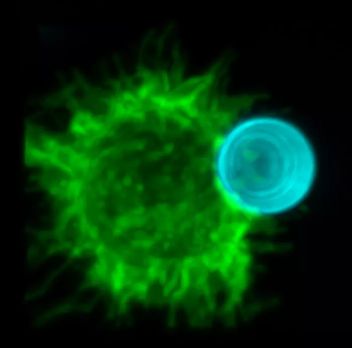
A cell glows bright against a black background. Like a flipbook, images meld into movement, showing an immune cell called a phagocyte approaching a dead cell, recognizing the dead cell as a potential danger, and ingesting its target through a process called phagocytosis.
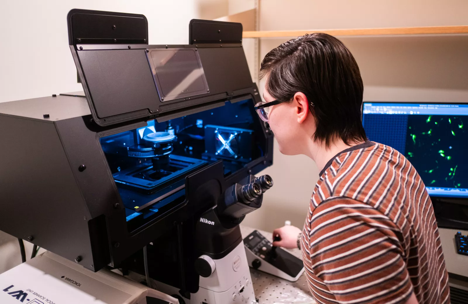
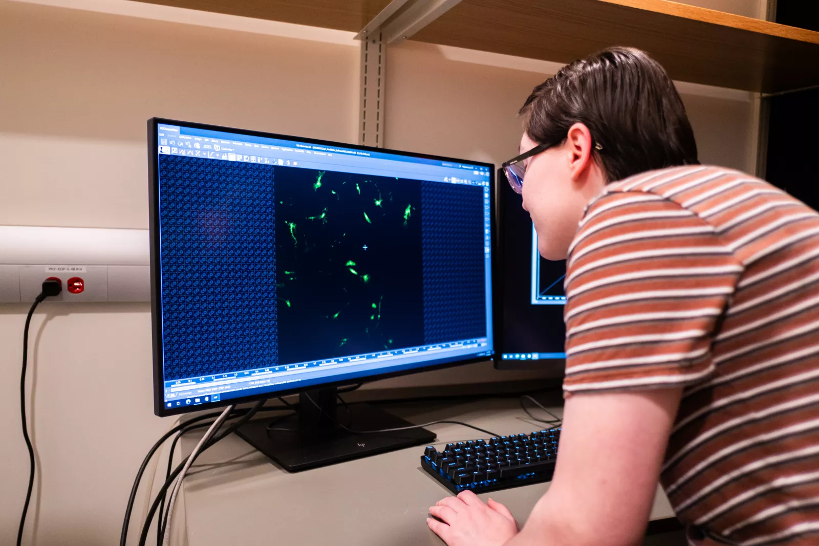
Phagocytosis often occurs within minutes. Being able to see that process moment-by-moment is now possible with the new spinning disk confocal microscope in the Park Science Building.
“What the microscope lets us do that we couldn't do before is capture three-dimensional snapshots of living phagocytes in action very quickly with minimal damage to the cells we are interested in learning about,” says Assistant Professor of Biology Adam Williamson.
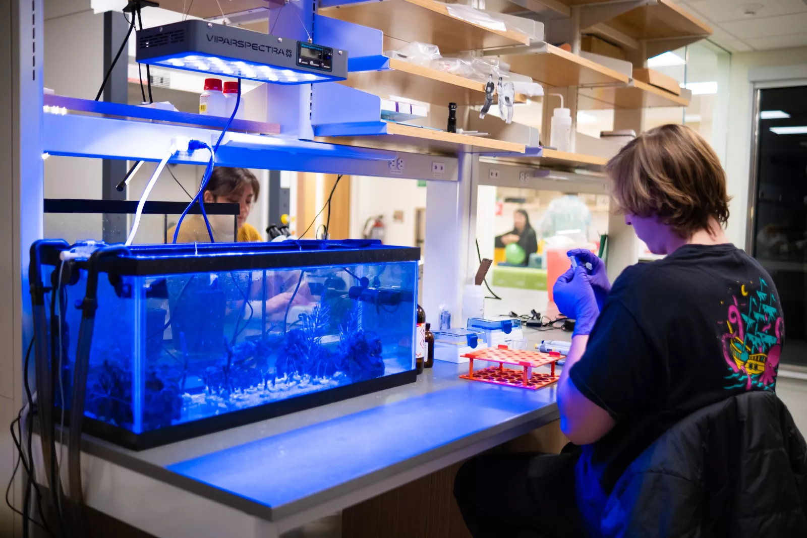
In addition to high-resolution imaging and the ability to capture real-time functions, the confocal microscope has a humidity- and temperature-controlled chamber that allows the scientists to work with living cells, something they could not do with an electron microscope. It is used by Williamson, Dean of Graduate Studies and Physics Professor May Cheng, and Assistant Professor of Physics Asja Radja, along with some of their students.
For Radja, the microscope was key to her joining the Bryn Mawr faculty in 2022 and being able to conduct her research. She studies patterns and shapes in biology, from those visible to the naked eye down to a microscopic scale, and the physics of how they are formed.
“The confocal allows us to look on a much smaller scale to understand what are the cellular mechanisms that could be leading to the emergence of macroscopic patterns, shapes, or shape changes and pattern changes,” she says.
Take, for instance, gorgonians, or soft corals. Unlike hard corals, Radja said, gorgonians seem to be resistant to the bleaching that is devastating coral reefs around the world. The corals house a colony of individual organisms called polyps that Radja is studying with Villanova master’s student Michael Drummond.
“It’s incredible,” Drummond says, adding that the microscope has given him a chance to see algae living on the polyps on a level he couldn’t before.
The first time Drummond used the microscope, he discovered the algae formed a unique pattern he had not seen in previous research. “He found something that no one ever published before,” says Radja. “That's really exciting.”
Before the college purchased the equipment, Williamson was using a confocal microscope located at the University of Delaware. It was a long distance for him to travel, and students couldn’t access a piece of equipment he says is “absolutely central to our research program.”
“This is a fundamental tool for asking biological questions, for asking biophysical questions,” he said, “and we’re grateful to have this piece of infrastructure at this institution that lets us do this work.”
Williamson is currently working with Kyle Bledsoe ‘24, majoring in biochemistry and molecular biology, on a research project involving the microscope. The project is funded by an NIH grant Williamson’s lab recently received in part because of access to the equipment.
"To be able to work with a student who might be new to science, who gets to walk into a microscopy room and see something that no one's ever seen before, I think that's incredibly exciting.”
– Assistant Professor of Biology Adam Williamson
Williamson’s research program’s focus on how immune cells recognize and fend off threats, as well as clean debris and dead cell through phagocytosis, dovetailed with Bledsoe’s interest in Batten Disease.
“It’s a group of genetic disorders that are called ‘lysosomal storage disorders,’” Bledsoe explains. “The lysosome is the part of the cell that takes up anything that's unwanted or needs to be degraded and uses enzymes to degrade all the kind of trash or debris in the cell.”
Batten disease primarily affects children and causes devastating symptoms, including visual impairment and loss of brain tissue, Williamson said, and it is usually fatal. The most common pediatric onset neurodegenerative disease, there are 14 different types of the disease, 13 of which have been mapped to a specific gene mutation.
Williamson and Bledsoe’s goal is to find out how, when working properly in cells, the genes mutated in Batten Disease protect the nervous system.
“We're hoping to offer some insight down the road for whether, from a cell biology perspective, we should be treating Batten disease as one disease that can all be treated the same way or as different diseases that might need different disease-specific treatments,” Williamson says.
Bledsoe is preparing human retinal cells for the study using CRISPR, a tool to edit genes. They then test the edits using PCR, which many people may have heard of as it relates to Covid tests, but here Bledsoe uses to amplify the DNA and verify the changes made through CRISPR.
They still have a lot of prep to do before their work is ready to go under the microscope, but they did train on it when it first arrived last summer.
“It's just an incredible machine,” Bledsoe says. “It’s very intricate and there's so many things that you're able to do with it, like looking at different wavelengths of light.”
With graduation approaching, Bledsoe’s time to research is running out, but they are confident other students will be excited to take over.
“I think there's something captivating about watching a cell carry out a process that when dysregulated has the potential to cause devastating disease,” Williamson says, “And for me as a mentor, to be able to work with a student who might be new to science, who gets to walk into a microscopy room and see something that no one's ever seen before, I think that's incredibly exciting.”